
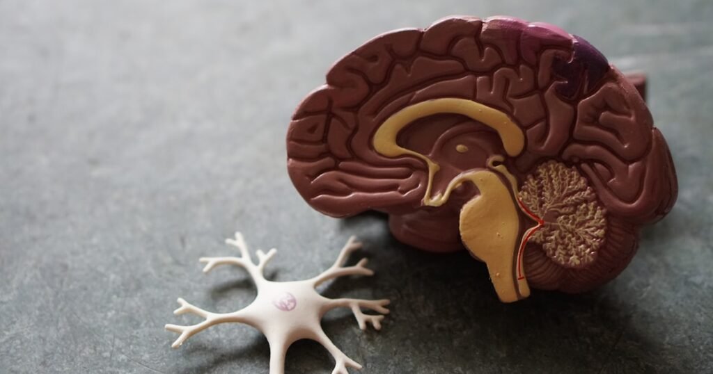
Whether caused by a fall, a sports collision, or a motor vehicle accident, a concussion is far more than a simple bump on the head. In the United States, roughly 2.8 million traumatic brain injuries occur annually, with mild traumatic brain injuries (mTBIs)—the clinical term for concussions—representing up to 90% of cases. Yet despite their prevalence, concussions remain widely misunderstood.
For decades, standard medical advice following a head injury was rigid: rest in a dark room, avoid all screens, conversation, and movement, and wait until all symptoms disappear. Today, clinical neuroscience has overturned this dogma. Major consensus guidelines—led by the 6th International Conference on Concussion in Sport (Amsterdam Consensus) and the National Institute of Neurological Disorders and Stroke (NINDS)—confirm that prolonged strict rest actually delays recovery, increases deconditioning, and worsens psychological distress. Adopting evidence-based active rehabilitation protocols can dramatically accelerate brain healing and prevent protracted disability.
Inside the Concussed Brain: The Neurometabolic Cascade
Understanding concussion recovery requires examining what occurs at the cellular level. When the head experiences sudden acceleration, deceleration, or rotational forces, the brain shifts within the cranial vault, stretching delicate axons and compromising cellular membranes.
This biomechanical stress triggers the neurometabolic cascade:
- Ionic Flux: Cellular membranes destabilize, allowing potassium ions to rush out of neurons while calcium floods in.
- Neurotransmitter Surge: Neurons indiscriminately release excitatory neurotransmitters, primarily glutamate, causing temporary neurological hyperexcitability.
- The Cellular Energy Crisis: Membrane pumps work overtime to restore ionic equilibrium, rapidly burning through cellular adenosine triphosphate (ATP) stores.
- Cerebrovascular Mismatch: Simultaneously, cerebral blood flow regulation becomes impaired, dropping by 20% to 50%. The brain demands surge precisely when microvascular fuel delivery is compromised.
Because structural CT and MRI scans evaluate gross anatomical integrity rather than cellular metabolism, they almost always appear normal following an uncomplicated concussion. The injury is functional, metabolic, and neurovascular rather than structural.
The Fall of “Cocoon Therapy”: Why Active Recovery Wins
The practice of prescribing prolonged sensory isolation—often called “cocoon therapy”—stemmed from a well-intentioned desire to protect the vulnerable brain. However, clinical trials published in the British Journal of Sports Medicine reveal that resting for more than 48 hours contributes to physical deconditioning, heightened autonomic dysregulation, and increased anxiety.
Modern clinical guidelines from the CDC and international concussion panels now recommend:
- Relative Rest (24 to 48 Hours): Brief cognitive and physical downtime immediately after the injury to let acute cellular volatility settle.
- Early, Gradual Activity: Beginning at 48 hours, patients are encouraged to resume light, symptom-limited cognitive and physical activities, such as brief walks around the home.
- Active Rehabilitation: Controlled physical movement acts as an active therapeutic agent rather than a risk to be avoided.
Sub-Symptom Threshold Aerobic Exercise
The cornerstone of modern concussion care is prescribed sub-symptom aerobic exercise. Researchers at the University of Buffalo established the Buffalo Concussion Treadmill Test (BCTT), which identifies the precise heart rate where concussion symptoms begin to rise.
Clinicians then prescribe 20 minutes of daily stationary cycling or walking at 80% to 90% of that threshold heart rate. Clinical trials show this targeted aerobic stimulation:
- Normalizes cerebral autoregulation, restoring healthy blood flow to healing brain tissue.
- Stimulates brain-derived neurotrophic factor (BDNF), a vital signaling protein supporting synaptic neuroplasticity, as highlighted in stroke rehabilitation and neuroplasticity guidelines.
- Recalibrates the autonomic nervous system, dampening persistent sympathetic hyperactivity.
Persistent Post-Concussive Symptoms (PPCS)
While most adults recover within two to four weeks, roughly 20% to 30% experience Persistent Post-Concussive Symptoms (PPCS) lasting months. When symptoms like headaches, brain fog, dizziness, and fatigue endure, they rarely reflect permanent brain damage. Instead, they typically arise from treatable secondary issues:
1. Vestibular and Oculomotor Dysfunction
Concussions often impair smooth ocular tracking, saccades, and convergence (how both eyes focus on near objects). This creates dizziness, visual strain, and motion sensitivity. Specialized vestibular and oculomotor therapy (VRT) retrains these gaze-stabilization reflexes, mirroring strategies used in vestibular migraine relief protocols.
2. Cervical Spine Whiplash
The force needed to cause a concussion far exceeds the threshold required to sprain the cervical spine. Damaged sensory receptors in the upper neck joints send aberrant signals to the brainstem, mimicking post-concussion headaches, brain fog, and dizziness. Targeted cervical physical therapy often eliminates symptoms that were assumed to be brain-based.
3. Post-Traumatic Migraines
Head trauma can sensitize the trigeminovascular system, transforming sporadic headaches into chronic post-traumatic migraines. Neurologists utilize targeted prophylactic treatments and neuromodulation, as detailed in research on nerve pain and post-traumatic migraine management.
Sleep, Nutrition, and Sensory Pacing
Supporting cellular repair requires optimizing daily metabolic foundations:
- Sleep Hygiene: Deep slow-wave sleep activates the brain’s glymphatic system, clearing metabolic waste and inflammatory byproducts, as explored in sleep architecture and brain recovery. Maintain strict, consistent sleep schedules without prolonged daytime naps.
- Anti-Inflammatory Nutrition: Polyphenol-rich whole foods and long-chain omega-3 fatty acids (DHA and EPA) provide essential structural lipids for neuronal membrane repair, as documented in clinical reviews of omega-3 fatty acids for neurovascular support.
- Cognitive Pacing: Practice structured sensory pacing rather than total screen bans. Work in 20-minute intervals followed by brief resting periods, gradually increasing tolerance.
Warning Signs: When to Seek Emergency Care
While most concussions follow an uncomplicated trajectory, head injuries can occasionally produce serious complications like intracranial hemorrhage. Seek immediate emergency evaluation if red flag signs emerge:
- A severe, progressively worsening headache.
- Repeated vomiting or persistent nausea.
- Unequal pupil sizes (anisocoria) or double vision.
- Difficulty waking up or marked lethargy.
- Slurred speech, limb weakness, or numbness.
- Convulsions, seizures, or escalating confusion.
Evidence Nuance and Clinical Perspective
Concussions are inherently heterogeneous injuries. Because much of the published literature originates from athletic cohorts, timelines must be adapted individually for older adults or individuals with pre-existing migraine conditions. Identifying an individual’s specific clinical profile—whether autonomic, cervical, vestibular, or ocular—remains essential for guided medical recovery.
Disclosure: This content is for informational purposes only and is not medical advice. Always consult a qualified healthcare provider before making changes to your health regimen.